Item 2 - Virtual biopsy in neurodegenerative diseases
The concept of virtual biopsy based on magnetic resonance imaging in neurodegenerative diseases is based on 3 facets.
- First, the use of radiomics analysis as an original method of image analysis to detect early morphological changes induced by neurodegeneration. Radiomics features include all the statistical measurements that can be calculated from the signal.
- First, the use of radiomics analysis as an original method of image analysis to detect early morphological changes induced by neurodegeneration. Radiomics features include all the statistical measurements that can be calculated from the signal.
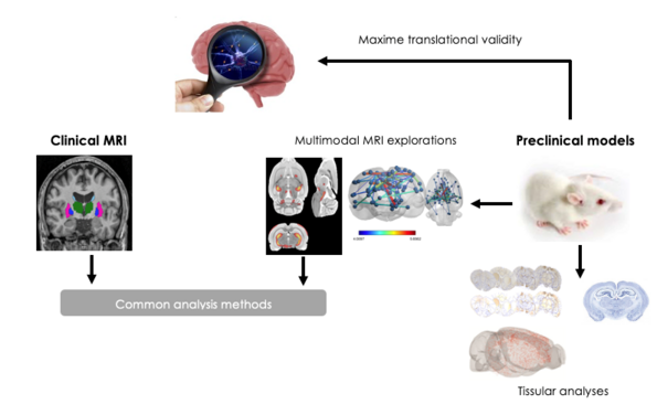
- And finally, the investigation of potential tissue signatures of these imaging characteristics by anatomo-radiological correlations using animal models. The first results of the method were obtained in the context of post-stroke cognitive decline (1) with international multicenter study validation (2).
Experiments have shown that certain radiomic features have higher sensitivity than traditional volumetric measurements when comparing them between patients with cognitive impairments and those without cognitive impairments. Combined with age, sex and simple cognitive scores, they predicted long-term cognitive deficit, while studying the tissue signature using the rat model of middle cerebral artery occlusion, mimicking a stroke, showed strong correlations between certain features measured in the hippocampus contralateral to the hippocampus. ischemic area and neuronal density.
The project was supported by the innovation prize from the University of Lille and a Franco-German PROCOPE grant. A patent is currently in progress and supported by INSERM-Transfert by a grant for the development of a proof of concept to facilitate industrial transfer.
Based on these results, the method was studied in other clinical situations, mainly in Parkinson's disease (PD) and showed very encouraging results on both aspects of the disease, motor (3) and non-motor (4). These results motivated the launch of a more ambitious project aimed at studying anatomo-behavioral-radiological correlations in different models of PD (doctoral thesis of Chirine Katrib, University of Lille, supported by a grant from the national charity France -Parkinson). The main results of this project highlight a link between signal changes captured by radiomic features and tissue changes such as neural death and protein aggregations (Publications under review).
- Betrouni N, Yasmina M, Bombois S, Pétrault M, Dondaine T, Lachaud C, Laloux C, Mendyk AM, Henon H, Bordet R. Texture Features of Magnetic Resonance Images: an Early Marker of Post-stroke Cognitive Impairment. Transl Stroke Res. 2020 Aug;11(4):643-652.
- Betrouni N, Jiang J, Duering M, Georgakis MK, Oestreich L, Sachdev PS, O'Sullivan M, Wright P, Lo JW, Bordet R; Stroke and Cognition (STROKOG) Collaboration. Texture Features of Magnetic Resonance Images Predict Poststroke Cognitive Impairment: Validation in a Multicenter Study. Stroke. 2022 ;53(11):3446-3454.
- Betrouni N, Moreau C, Rolland AS, Carrière N, Chupin M, Kuchcinski G, Lopes R, Viard R, Defebvre L, Devos D. Texture-based markers from structural imaging correlate with motor handicap in Parkinson's disease. Sci Rep. 2021 1;11(1):2724.
- Betrouni N, Lopes R, Defebvre L, Leentjens AFG, Dujardin K. Texture features of magnetic resonance images: A marker of slight cognitive deficits in Parkinson's disease. Mov Disord. 2020 Mar;35(3):486-494.